What it does
Training Simulator that replicates the anatomy and mechanics of the human eye, allowing ophthalmology students and residents to practice surgical techniques in realistic conditions, improving precision, confidence, and readiness for real procedures.
Your inspiration
The idea came after a conversation with a ophthalmologist who shared his concern about the lack of realistic training tools in Local eviroment. Hearing that future surgeons were learning with improvised methods made me realize the responsibility we have to improve this. I felt driven to create a solution that could offer precise, safer and more accessible training, because every patient deserves a high quality surgery made by a well prepared surgeon.
How it works
The Ophthalmic Surgery Training Device features a mechanical system that simulates the resistance of the extraocular muscles, providing tactile feedback that mimics the force dynamics encountered during real eye surgeries. The facial structure is anatomically shaped, allowing trainees to identify and use proper support points, which are essential for stability during procedures. The device uses varied materials: a flexible surface in the ocular cavity enables realistic eyelid manipulation with surgical tools, while the rest of the face is made of a more rigid material to simulate facial firmness. The eye model remains stable during use, and the entire device can be securely fixed to a working surface. Additionally, it includes a drainage system to manage surgical fluids during practice, enhancing hygiene and realism. The simulator is also fully disassemblable, allowing for easy cleaning and maintenance between training sessions.
Design process
The design process began by analyzing the basic models used by ophthalmology students to practice surgical techniques. After three validation sessions with a professional ophthalmologist, we identified key limitations in existing training tools and defined the real requirements for a more effective solution. The problem was then broken down into smaller challenges, starting with the development of a mechanism to replicate the natural resistance of extraocular muscles. Several digital models were created and tested through medium-fidelity prototypes, which were evaluated by the ophthalmologist to refine the design through iterative feedback. Once the mechanism was defined, new challenges emerged—such as designing a housing that could simulate the human face. This phase began with reverse engineering a 3D scan of a real face and evolved into a multi-material shell capable of replicating facial morphology and draining surgical fluids. Over ten low- and medium-fidelity prototypes were developed using digital fabrication methods like resin and filament 3D printing, allowing the device to aim high anatomical precision and mechanical accuracy.
How it is different
What truly sets this device apart is its thoughtful combination of features rarely found together in a single, low-cost solution: realistic eyelid manipulation enabled by flexible materials in the ocular cavity, facial rigidity for accurate hand positioning, a built-in drainage system for surgical fluids, and a fully disassemblable design for easy cleaning and maintenance. To ensure high fidelity and precision, the simulator was developed using digital fabrication techniques—primarily 3D printing in resin and filament—allowing for rapid prototyping and anatomical accuracy. Recognized with a national invention patent in Colombia, this device is more than a training tool—it’s a step toward democratizing access to high-quality surgical education. By combining innovation in health and technology, it empowers future ophthalmologists to train safely, confidently, and affordably.
Future plans
The device is currently in an early stage of transition toward commercial viability. After securing a patent for the invention in Colombia, the next major challenge is to meet the quality and production standards required to address the needs of the local market. This involves carefully planning a sustainable business model, mapping the target market, and identifying its size and specific interests. The goal is to build a potential buyers network and position the device as the preferred simulator for educational institutions and medical training centers, thanks to its accessibility, versatility, and low cost.
Awards
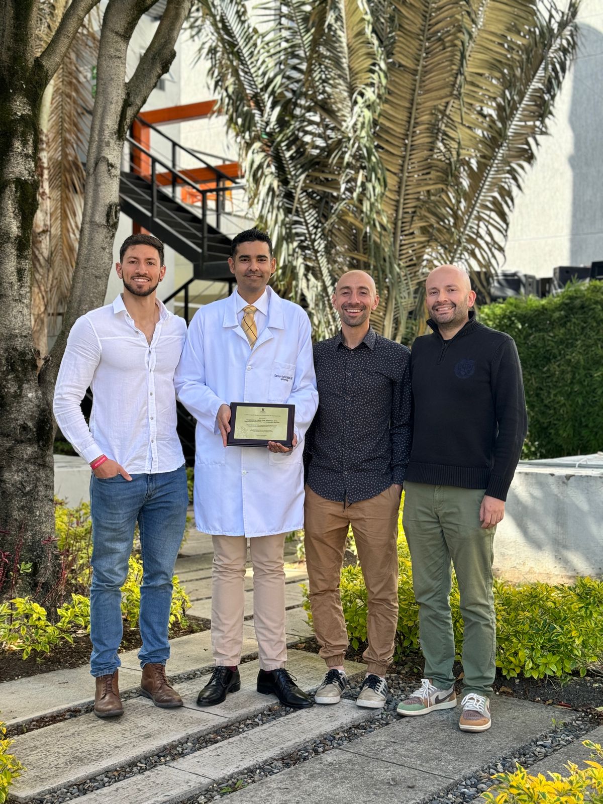
This invention was officially recognized with a national patent granted in Colombia on October 30, 2021, under the title “DISPOSITIVO DE ENTRENAMIENTO PARA CIRUGÍA OCULAR.” The patent acknowledges the originality and technical contribution of the simulator to the field of surgical education. N° NC2021/0014712.
Created by
- Luis Alejandro Estrada Rodriguez
- My website
University / Institution
- Universidad El Bosque
Region
- Colombia


Share this page on